Abstract
This pilot study investigated whether short-term exposure to natural, untreated rock crystal is associated with measurable changes in human biophoton emission, as recorded using non-invasive Bio-Well technology. Two pilot experiments were conducted. In the first experiment, 120 participants were assessed before and after holding a rock crystal for 10 minutes. In the second experiment, 28 participants were assessed across a baseline-placebo-rock crystal sequence to evaluate placebo-related effects and responses associated with rock crystal exposure. Data were analysed using descriptive statistics, the Wilcoxon signed-rank test, and Spearman’s correlation analysis. Rock crystal exposure was associated with statistically significant changes in Bio-Well parameters, particularly total recorded energy and indicators related to the head, cerebral cortex, and cardiovascular system. The findings also suggested that responses may vary according to age, with a reduction in stress-related indicators observed in the older participant group. The placebo study showed that both glass and rock crystal produced similar changes, possibly owing to their similar chemical composition. Overall, this study provides preliminary quantitative data in an area where previous evidence has largely been empirical or qualitative. The findings suggest that responses to rock crystal exposure can be objectively recorded using non-invasive biophysical methods and support further investigation into crystal therapy and its potential mechanisms of action.
1. Introduction
The use of minerals and crystals has accompanied human cultures for millennia, including in Ancient Egypt, Mesopotamia, the Indus Valley civilisation, and other ancient cultures worldwide. In these contexts, crystals were used in magical rituals and healing practices, as reflected, for example, in the Egyptian Ebers Papyrus and in the Assyrian-Babylonian Abnu Shikinshu texts. Historical associations between crystals and their presumed effects are also reflected in the names of certain crystals. Across different regions and cultural traditions, some crystal names appear to indicate an assumed direction of action. For instance, the term nephrite is derived from the Greek nephros, meaning “kidney,” thereby reflecting a historical association between this crystal and the kidneys. Hippocrates, the ancient Greek physician often regarded as the father of modern medicine, also referred to the use of stones in the treatment of certain diseases [1]. The Romans later adopted and developed these traditions, using stones both for medical purposes and as ornaments [2].
In Eastern cultures, particularly in the Altai and Himalayan regions, it has been – and in some contexts remains – customary to use mixtures of powdered stones that are manually ground for this purpose in healing practices [3-6]. These mixtures were prescribed by a master to be taken at a specified time, accompanied by prayer.
One of the most notable historical figures associated with the therapeutic use of stones and the early traditions of crystal therapy was Hildegard von Bingen (1098–1179), a Benedictine abbess, polymath, mystic, and author of medical writings who lived in medieval Germany. In her Physica (Liber subtilitatum diversarum naturarum creaturarum), a work belonging to the medieval tradition of natural philosophy and medicine, Hildegard described the properties of various natural substances, including stones, their effects on humans, and their possible therapeutic applications [7]. These ideas were later revisited by Michael Gienger in Die Heilsteine der Hildegard von Bingen, in which Hildegard’s descriptions of healing stones were interpreted and systematised in the context of modern crystal therapy [8].
Until the Renaissance, various natural substances, including crystals, formed an integral part of healing practices. However, during the seventeenth and eighteenth centuries, with the development of scientific medicine and pharmacology, medical practice gradually shifted towards chemically defined and experimentally based therapeutic approaches [9]. As a result, traditional healing practices, including the use of crystals, were increasingly subjected to critical evaluation and regarded as insufficiently supported by scientific evidence.
Jane Ann Dow (1936-2008) is often recognised as a significant early figure in the revival of modern crystal therapy. Through her work, the tradition of healing with crystals began to re-emerge after several centuries of interruption. Dow emphasised that the human being should not be understood merely as a physical body, but as a complex and integrated system. She believed that the medicine of the future would address the human being as an integrated whole [10].
Another major contributor to the formation of modern crystal therapy was Michael Gienger (1964-2014), whose work focused on linking the practical use of crystals with their mineralogical properties. He was among the first authors to present empirical observations on the application of crystals in humans and to interpret their effects in relation to each crystal’s mineralogical profile, including its physical and chemical characteristics. According to Gienger, the nature of a crystal’s effects may be influenced by several basic factors: geological formation conditions, chemical composition, crystalline structure, including crystal system, and colour [11-13]. In 1995, he founded the International Research Organisation for the Healing Properties of Stones (Forschung und Entwicklung der Steinheilkunde; SHK-Forschung), where long-term practical observations were collected, organised, and examined in relation to mineralogical properties. The organisation remained highly active until his death.
Gienger’s work was directly related to the development of crystal therapy in Lithuania. In the same year, 1995, Audronė Ilgevičienė founded the Pars Fortunae Astromineralogy Centre. After 2000, Ilgevičienė continued empirical explorations in collaboration with Gienger. This active and intensive work attracted individuals from various professional backgrounds, who studied, practised, and participated in these explorations. In 2017, on Ilgevičienė’s initiative, the Association of Lithotherapy was established, bringing together people from different professions who applied crystals for their own health and well-being. Thus, after nearly three decades of continuous and intensive practical work in this field, a more refined system of lithotherapy, characterised by specific instructions and detailed explanations, was developed. This long-term work contributed to the transformation of crystal therapy into a more coherent and methodical system for the application of crystals to human physical, emotional, mental, and spiritual health [14-17].
The effects of crystals on humans are viewed controversially in contemporary society. Much of the available information is subjective and often insufficiently substantiated, creating conditions for misuse and for the dissemination of misleading claims in this field. This contributes to a superficial and consumer-oriented understanding of the effects of crystals on humans and may discredit a practice with a long historical presence in human cultures. Because of the limited scientific substantiation and the widespread availability of subjective and superficial information online, two opposing extremes have emerged: the use of crystal therapy as a purported remedy for all diseases, and the complete dismissal or denial of the field. Both extremes hinder a more objective consideration of the possibility that crystals, like plants or other natural objects, may exert certain effects. At present, scientific uncertainty remains regarding the effects of crystals, which we seek to investigate and address using currently available methods.
With the recent emergence of technologies aimed at integrally assessing the state of the human organism, such as Kirlian photography, it has become possible to evaluate the effects of crystals on humans in a more objective manner. For this purpose, we used Bio-Well technology, developed by K. Korotkov, which has been widely applied internationally [18], [19]. Gas discharge visualisation, also referred to as electrophotonic imaging, is a non-invasive biophysical method used to record stimulated optical emission from human tissues. It involves the application of a high-voltage pulsed electric field, which induces corona discharge signals at the skin surface. These signals are captured optically and subsequently analysed using dedicated software [18]. The analysis of the obtained signals is interpreted in relation to the general functional state of the organism [19].
Within biophoton emission research, the 1988 publication by Fritz-Albert Popp and co-authors is regarded as one of the early systematic reviews of the field. The paper summarised the evidence then available on ultra-weak spontaneous photon emission in biological tissues and organisms, helped standardise terminology by promoting the concept of “biophoton emission,” and proposed theoretical interpretations linking this phenomenon to biological processes [20]. It has since been frequently cited as an important reference point for the further experimental and theoretical development of the field [21], [22].
This methodology has been reported to be sufficiently sensitive for investigating several domains, including psychological state, autonomic nervous system activity, and stress levels. Previous studies have reported correlations between Bio-Well parameters and physiological indicators such as heart rate variability, blood pressure, and other markers related to autonomic nervous system function [23]. The method has also been applied in studies assessing the effects of therapeutic interventions, including osteopathic treatment [24]. In addition, Bio-Well-derived parameters have been reported to be associated with metabolic and clinical indicators, such as blood glucose levels and general functional reserve [25]. Efforts have also been made to establish normative values for this methodology across different populations, suggesting its increasing application in scientific research [26].
Despite the increasing use of Bio-Well technology in studies of psychophysiological and functional states, quantitative research examining the effects of natural crystals using this method remains very limited. The novelty of the present study lies in its attempt to provide preliminary quantitative evidence on short-term exposure to natural, untreated rock crystal using a non-invasive biophysical assessment method. Therefore, this study aimed to assess whether short-term exposure to rock crystal is associated with measurable changes in Bio-Well parameters, including total recorded energy, stress-related indicators, and selected system-specific projection indices.
2. Methods
Study design. Two pilot studies were conducted. The first study included 120 participants: 99 women with a mean age of 45.29±7.16 years and 21 men with a mean age of 47.54±11.76 years. The second study examined responses to a non-crystalline structure in the placebo group and included 28 women with a mean age of 47.19±10.55 years.
In the first pilot study, participants were assessed in a seated position. After entering the study room, each participant sat quietly for 10 minutes, after which biophoton emission was recorded. The participant then held a rock crystal, a colourless variety of quartz weighing 300 g, in the left hand for 10 minutes while remaining seated (Fig. 1). Biophoton emission was then recorded again. Rock crystal was selected for the study because of properties empirically attributed to it, particularly its presumed energetic effect, which may be associated with relatively rapid changes in biophoton emission. The aim of this study was to evaluate objectively the effect of natural rock crystal on biophoton emission. Although the study was non-invasive, each participant completed a voluntary informed consent form.
In the second study, only women were included. First, baseline biophoton emission was recorded. Each participant was then given a small pouch and told that it contained a stone; however, the pouch in fact contained a glass object intended to imitate a natural crystal. After holding the pouch for 10 minutes, biophoton emission was recorded for a second time. The first pouch was then replaced with a second one, which the participant was told likewise contained a stone; in this case, the pouch contained a natural, untreated quartz crystal, namely rock crystal. After holding the second pouch for 10 minutes, biophoton emission was recorded for a third time, completing the baseline–placebo–crystal sequence. Participants were not informed about the actual contents of either pouch.
It should be noted that both rock crystal and glass are composed of SiO₂ and therefore share the same chemical composition. However, they differ in their formation conditions and structural organisation. Glass is a human-made material of non-natural origin with an amorphous structure. In contrast, rock crystal is a naturally occurring mineral with a trigonal crystal system, meaning that its constituent particles are arranged in an ordered, symmetrical structure. According to CIBJO standards, a natural untreated mineral is defined by its origin and formation process – specifically, as a material formed in nature through geological processes without human intervention [27]. A natural, untreated rock crystal was used in the present study.
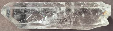
Rock crystal is a colourless variety of macrocrystalline quartz that belongs to the oxide mineral class. Quartz is one of the most abundant minerals in the Earth’s crust, with a hardness of 7 on the Mohs scale. The crystal used in the study is shown in Fig. 1.
Fig. 1Rock crystal used in the study. Dimensions: 150×40 mm; weight: 323 g
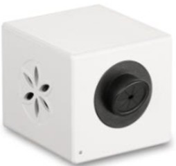
Measurements were performed using the Bio-Well device. The scanning procedure is rapid, simple, and non-invasive. According to the manufacturer’s documentation, the device is registered with the FDA (registration No. 3014299556) and holds EU and UL certifications. The device used for recording is shown in Fig. 2.
Fig. 2Bio-Well recording equipment
To reduce immediate physiological variability, all participants were assessed in a seated position after a 10-minute resting period before baseline recording, and all measurements were performed using the same Bio-Well device and a standardized recording procedure. Baseline and post-exposure measurements were conducted consecutively within the same session, thereby minimizing short-term variation related to the time of day and environmental conditions. However, factors such as prior physical activity, caffeine intake, acute emotional state, sleep quality, and individual differences in skin conductivity were not systematically recorded or controlled. These factors are therefore acknowledged as potential confounders and should be controlled more rigorously in future studies.
The data were analysed using SPSS software. Descriptive statistics were used to summarise the main Bio-Well parameters, including the stress index, total recorded energy, organ system imbalance, and selected organ-specific energy indicators. Results are presented as mean ± standard deviation (M ± SD). As measurements were obtained from the same participants before and after exposure, the data were treated as paired observations. Because the distributions of the investigated variables could not be assumed to follow a normal distribution, the Wilcoxon signed-rank test was applied to assess changes between baseline and post-exposure measurements. Separate analyses were conducted for women and men; the female group was further divided by median age into younger and older subgroups. Spearman’s rank correlation was used to examine relationships between the investigated parameters, including stress, total energy, organ imbalance, and energy indicators of selected organ systems. Statistical significance was set at 0.05.
3. Results
First, we aimed to determine whether rock crystal exposure was associated with any measurable response. Women and men were analysed separately; the selected parameters are presented in Table 1. Although the Bio-Well system provides 1,496 derived parameters, this article focuses on several key indicators: changes in stress level, total biophoton emission intensity (expressed as “Energy” [J×10⁻2]), and the overall imbalance of recorded emission across different organ systems. Since such a subtle exposure may produce sex-specific responses, the study sample was divided into a female group ( 99) and a male group ( 21). The responses observed in both groups during the first study are shown in Table 1. Owing to the small number of male participants and the relatively weak observed effect, the male group was not included in further detailed analysis, as the reliability of these results was considered limited.
Table 1Comparison of selected Bio-Well parameters at baseline and after holding a crystal in the left hand. Data are presented as mean ± standard deviation (M±SD). Statistically significant differences are shown in bold
Parameter | Female, 99 | Male, 21 | |
1 | Stress, before | 3.33±1.02 | 3.19±0.51 |
Stress, after | 3.29±1.02 | 3.41±0.12 | |
2 | Energy, before | 45.48±8.61 | 48.1±6.65 |
Energy, after | 46.55±6.84 | 46.21±7.6 | |
3 | Imbalance, before | –2.37±12.57 | 0.84±5.34 |
Imbalance, after | 1.29±9.58 | 3.33±8.2 |
To examine the effect of the exposure on women of different ages in greater detail, the female group was divided into two subgroups by median age: a younger group (aged < 47 years) and an older group (aged > 47 years). Changes in the investigated parameters are presented in Table 2. Statistically significant differences between baseline and post-exposure measurements are shown in bold.
A tendency towards an increase in the stress index was observed in the younger women’s group, whereas a decrease was observed in the older group. The energy index increased in both groups.
Given that the exposure applied in this study was of very low intensity, different organ systems were expected to respond differently. Upon reviewing the full list of obtained parameters, we observed that changes in certain systems were particularly pronounced. The systems showing the most marked and statistically reliable responses were selected for further analysis and are highlighted in bold.
Table 2Bio-Well parameters in women of different age groups before and after holding a crystal in the left hand. Data are presented as mean ± standard deviation (M ± SD). Statistically significant differences are shown in bold
Parameter | Female, (Age < 47 yr) 50 | Female, (Age > 47) 47 | |
1 | Stress, before | 3.32±0.87 | 3.35±1.17 |
Stress, after | 3.41±1.16 | 3.17±0.85 | |
2 | Energy, before | 46.33±8.12 | 44.55±9.08 |
Energy, after | 46.98±6.64 | 46.12±7.08 | |
3 | Imbalance, before | –1.96±9.44 | -2.78±2.17 |
Imbalance, after | 0.39±8.89 | 2.21±1.46 |
Table 3Changes in selected organ-specific energy parameters before and after exposure in female participants during the first study (N= 99). Data are presented as mean ± standard deviation (M±SD)
Organ-specific energy parameters | Before | After | p-value | |
1 | Head | 4.1071 ± 0.84354 | 4.2609 ± 0.71705 | 0.024 |
2 | Cortex | 3.8977 ± 0.09348 | 4.0836 ± 0.08804 | 0.014 |
3 | Cardiovascular system | 4.3532 ± 0.09601 | 4.4892 ± 0.07776 | 0.027 |
In the female group, more pronounced and statistically significant changes were observed in energy parameters associated with the brain and cardiovascular system.
In the second experiment, designed to assess the placebo effect, a significant change in the biophoton energy index was observed (Table 4). The absence of significant changes in the other parameters may be attributed to the relatively small sample size in the second study.
Table 4Changes in selected Bio-Well parameters across the baseline-placebo-crystal sequence in female participants during the second study (N= 28). Data are presented as mean ± standard deviation (M±SD)
Parameters, 28, female | Mean | Std. Deviation | p-value compared with baseline (before) |
Stress_Before | 2.8004 | 0.35048 | |
Stress_Placebo | 2.8439 | 0.40549 | n.s. |
Stress_Crystal | 2.7868 | 0.36950 | n.s. |
Energy_Before | 53.2850 | 5.38380 | |
Energy_Placebo | 54.4557 | 4.94911 | 0.053 |
Energy_Crystal | 55.0268 | 4.79498 | 0.010 |
Organ imbalance. % Before | 2.7532 | 8.11872 | |
Organ imbalance. % Placebo | 0.1621 | 6.45422 | n.s. |
Organ imbalance. % Crystal | 0.3707 | 8.14471 | n.s. |
However, when changes in individual systems were examined during the second study, clear and statistically significant changes were observed in the energy indices associated with the head, cerebral cortex, and cardiovascular system – both following exposure to the rock crystal and following exposure to the placebo condition (Table 5). This finding was initially unexpected; however, it may be explained by the fact that both the crystal and the placebo material were composed of the same chemical substance, SiO2. Accordingly, similar changes were observed in both conditions.
Relationships between the investigated variables were further examined using Spearman’s correlation analysis. The analysed system-specific energy parameters showed significant correlations with the overall energy index. The energy indices associated with the brain and cardiovascular system were also significantly correlated with each other. In contrast, the kidney energy index did not correlate significantly with the other system-related parameters. A negative correlation was observed between the stress index and system-specific energy indices, suggesting that higher stress levels were associated with lower energy values across these systems.
In the first study, similar correlation patterns were observed across baseline and post-exposure measurements in the female group ( 99) (Table 7).
Table 5Organ-specific energy parameters during the second study in female participants (N= 28). Data are presented as mean ± standard deviation (M±SD)
Organ-specific energy parameters, N = 28, female | Mean | Std. Deviation | P value compared with baseline (before) |
Head_Before | 4.6261 | 0.68267 | |
Head _Placebo | 4.8614 | 0.69571 | 0.014 |
Head _Crystal | 4.9154 | 0.82394 | 0.013 |
Cortex_Before | 4.3154 | 0.60924 | |
Cortex _Placebo | 4.6861 | 0.77848 | 0.003 |
Cortex _Crystal | 4.8054 | 0.68788 | 0.000 |
Cardiovascular system_Before | 4.8621 | 0.61807 | |
Cardiovascular system_Placebo | 5.0775 | 0.67618 | 0.031 |
Cardiovascular system_Crystal | 5.1368 | 0.67591 | 0.003 |
Table 6Spearman correlations between selected Bio-Well parameters at baseline. Statistically significant correlations are shown in bold
Stress_B | Energy_B | Organ imbalance_B | Head_B | Cardiovascular system_B | Kidneys_B | |
Stress_B | 1.00 | –0.62 | –0.23 | –0.62 | –0.61 | –0.34 |
Energy_B | –0.62 | 1.00 | 0.17 | 0.86 | 0.85 | 0.69 |
Organ imbalance_B | –0.23 | 0.17 | 1.00 | 0.10 | 0.07 | 0.21 |
Head_B | –0.62 | 0.86 | 0.10 | 1.00 | 0.83 | 0.39 |
Cardiovascular system_B | –0.61 | 0.85 | 0.07 | 0.83 | 1.00 | 0.44 |
Kidneys_B | –0.34 | 0.69 | 0.21 | 0.39 | 0.44 | 1.00 |
Table 7Spearman correlations between baseline and post-exposure Bio-Well parameters in female participants during the first study (N= 99)
Stress_A | Energy_A | Organ imbalance_A | Head_A | Cardiovascular system_A | Kidneys_A | |
Stress_B | 0.78 | –0.52 | 0.13 | –0.54 | –0.51 | –0.25 |
Energy_B | –0.46 | 0.75 | –0.15 | 0.70 | 0.68 | 0.51 |
Organ imbalance_B | –0.10 | –0.04 | 0.14 | –0.02 | –0.05 | 0.02 |
Head_B | –0.49 | 0.64 | –0.10 | 0.76 | 0.66 | 0.27 |
Cardiovascular system_B | –0.48 | 0.64 | –0.16 | 0.65 | 0.74 | 0.35 |
Kidneys_B | –0.17 | 0.47 | –0.08 | 0.29 | 0.30 | 0.54 |
Significant correlations were observed between the overall energy index and the energy indices associated with the brain and cardiovascular system. In contrast, the associations involving the kidney energy index showed a somewhat different pattern.
The results of the first study were further analysed using the Wilcoxon signed-rank test. The statistical problem addressed in this analysis and the method used to evaluate it are described below.
The variables were defined as follows: – the value of the investigated parameter before the experiment; – the value of the parameter after the experiment (after crystal exposure).
To assess the effect of crystal exposure on a given parameter, a group of participants was studied. The characteristic of interest was measured before and after exposure, yielding paired samples and . Since the same participants were measured at both time points, the data consisted of paired observations. As the assumptions required for Student’s paired -test were not met, the Wilcoxon signed-rank test was selected to compare the pre- and post-exposure values:
The difference was calculated for each pair of observations: .
Where 0, the value is excluded from further calculations, and the effective sample size is taken as the number of non-zero values. The absolute values are then ranked. Ranks corresponding to negative values are termed negative ranks, and those corresponding to positive values are termed positive ranks. The sum of positive ranks is denoted . For small samples ( 25), tables of critical values of the test have been compiled. For larger samples, the distribution of is approximated by the standard normal distribution:
where:
The -value is given by: , where denotes a standard normal random variable.
The statistical analysis of the experimental data was then carried out. As the experiments generated a large number of organism-specific parameters, it was not possible to examine all of them in detail within the scope of this article; only selected results are therefore presented.
Table 8Statistically significant changes in the investigated parameters during the first study, according to the Wilcoxon signed-rank test
No. | Parameter | Wilcoxon signed-rank test p- |
1. | Organ imbalance % | 0.00447 |
2. | Male stress | 0.03115 |
3. | Male kidneys | 0.04001 |
4. | Female organ imbalance % | 0.01725 |
5. | Female head | 0.0482 |
6. | Female (age > 47 years) organ imbalance % | 0.02688 |
As shown in Table 8, the -value of the Wilcoxon signed-rank test is below 0.05 for each listed parameter; accordingly, is rejected in every case. These results indicate statistically significant differences between the pre- and post-exposure measurements for the parameters listed.
4. Discussion
The findings suggest that short-term exposure to rock crystal may be associated with small but statistically significant changes in biophoton emission parameters, which may be explained by the subtle effect of crystals on the human organism. To the best of the authors' knowledge, this study may be among the first to report the statistical significance of such associations.
All living organisms emit spontaneous ultra-weak light radiation, estimated at approximately 102-103 photons per second per square centimetre, thought to originate from excited electronic states generated during oxidative metabolic processes [22]. In the present study, the Bio-Well system was used to assess parameters related to this type of emission.
Bio-Well technology analyses gas discharge images using a projection model based on acupuncture points and meridians. Within this model, derived parameters are interpreted as reflecting functional aspects of different organ systems. The technology thus provides an indirect assessment of the organism’s functional state [18], [28]. In the present study, a statistically significant increase was observed in Bio-Well-recorded signal intensity associated with projection indicators of the cerebral cortex, head, and cardiovascular system. Researchers who have qualitatively examined the effects of crystals on the human organism have previously observed that crystals appear to act first and most prominently on cognitive functions and perception, followed by effects on emotions and, subsequently, on the physical body [11]-[17]. Another noteworthy observation was the statistically significant response of the cardiovascular system, which may be interpreted in the context of the close brain–heart relationship described in neurocardiology studies by researchers at Oxford [29] and further explored by Rollin McCraty and the HeartMath Institute [30], [31].
Although Bio-Well technology continues to be viewed critically with regard to measurement reliability and the interpretation of results, the growing body of research accumulated in recent years suggests its potential for analysing various physiological and psychophysiological states and their changes. For example, a systematic review of 136 publications – comprising 26 systematic reviews, 19 randomised controlled trials, 18 case reports or case series, and 13 cohort studies – reported that the gas discharge visualisation (GDV) method is an easy-to-use and versatile diagnostic tool. This method is applicable to the assessment of treatment-related changes (before and after therapeutic procedures), the monitoring of emotional and somatic states, and the evaluation of functional changes in the organism [23]. A study comparing 56 individuals with colorectal cancer with healthy controls found that GDV parameters differed substantially between participants with colorectal neoplasia and healthy individuals, suggesting that GDV technology may have potential as an auxiliary screening tool in diagnostics [32].
A further systematic review, which analysed 42 studies, demonstrated the potential of the GDV method for identifying functional deviations from normative patterns in certain bodily systems, particularly in relation to endocrine and immune system disturbances. GDV has also been used to assess well-being in healthy individuals and to monitor the effects of interventions including exercise, meditation, acupuncture, music therapy, and massage [33]. Overall, studies using the GDV method have demonstrated statistically significant differences between healthy and pathological groups in parameters such as emission area, intensity, and entropy.
A prospective study of 200 individuals with neuropathic pain found that GDV parameters were significantly correlated with psychological stress but showed little association with the intensity of somatic or nociceptive pain [34]. These findings suggest that GDV parameters may serve as a relatively specific biomarker for anxiety or psychological stress.
From the perspective of alternative medicine, the human body is regarded as a complex system comprising not only the physical body but also a biofield system – defined as a complex network of electromagnetic, quantum, and biochemical interactions [19], [35]. Within this model, disease is conceptualised as a disturbance of energetic balance that may eventually manifest as physical symptoms. Several approaches within alternative medicine, including acupuncture, homeopathy, and crystal therapy, have been classified as forms of “resonance medicine.” In this context, crystals are described as structurally stable substances capable of interacting with the human energy field through resonance principles [36]. Experimental studies have shown that living systems are sensitive to extremely low-intensity signals and that cellular processes can be modulated by electromagnetic fields [37]. As the human organism may be regarded as an integrated energetic system sensitive to external frequencies and vibrations [38], the interaction between crystals and the human energetic system has been associated with the maintenance of psychophysiological balance and the activation of natural self-regulatory processes.
The effects of crystals on the human organism may also be considered within the framework of quantum processes, particularly with regard to possible interactions with microtubules in neurons and other cells. The crystalline lattice of a crystal, determined by the mass and charge of its constituent atoms, constitutes a specific structural organisation that may interact with quantum fields. Such structures have been proposed to generate quantum and electromagnetic oscillations, including zero-point fluctuations. In proximity to biological tissues, these oscillatory processes could potentially influence cellular activity by engaging sensitive molecular mechanisms. Based on the work of Stuart Hameroff [39-42], microtubules – which are widely distributed in cells, particularly in neurons – may represent one possible site of such interaction, with initial effects potentially occurring at the level of tubulin proteins (alpha- and beta-tubulin) through changes in quantum states and electron distribution. Related concepts are discussed in the work of Roger Penrose concerning quantum processes at the microtubular level [43], [44]. Such changes could theoretically initiate weak photon emission, which may in turn influence mitochondrial function. Activated mitochondria may then emit biophotons, contributing to signal transmission within the cell and to surrounding tissues via the primary vascular system or the intercellular medium [22]. Information transfer may additionally occur through soliton-like electrical impulses, potentially contributing to the coordination of intercellular responses [45].
It may therefore be hypothesised that the effects of crystals are mediated through various mechanisms described in quantum physics and biophysics. It is important to note, however, that several of these pathways have not yet been conclusively demonstrated and are unlikely to represent the only mechanisms involved; alternative routes of interaction – for example, through DNA molecules, proteins, cell membranes, or electromagnetic fields – may also exist. A more detailed investigation of these processes is currently constrained by available technological capabilities, and further interdisciplinary research is needed.
The present study extends the assessment of human-environment interactions and points to new possibilities for conceptualizing such interactions in the context of human health. This may be regarded as a further step forward in the investigation of these relationships, in line with broader developments in contemporary science discussed by Krauss [46].
In summary, the findings of this study provide data indicating that crystals influence human biophoton emission. These results should be interpreted with caution. One important limitation of this pilot study is that several factors potentially affecting Bio-Well readings, including prior physical activity, caffeine intake, acute emotional state, sleep quality, and individual differences in skin conductivity, were not systematically recorded or controlled. Future studies should incorporate stricter methodological controls, larger sample sizes, and systematic investigation of the underlying mechanisms in order to establish the biological basis and practical significance of these phenomena. The effects observed in this study were relatively small, which may reflect the low intensity of the exposure, the limited sample size – particularly in the male group – and substantial individual variability, all of which are characteristic of early-stage pilot research and underscore the need for larger, more rigorously controlled experiments.
5. Conclusions
1) The statistically significant changes in biophoton emission observed in this study suggest that the effects of crystals on the human body can be objectively recorded using non-invasive biophysical methods.
2) Responses to crystal exposure appear to be individualised, with differences observed among participants, including age- and sex-related response patterns.
3) The data indicate that the effects of crystals are not uniform and are associated with specific changes in distinct bodily systems, particularly in the cerebral and cardiovascular domains.
4) The results of the placebo-controlled experiment indicate that some of the observed effects may be attributed to nonspecific or psychophysiological mechanisms; future studies should therefore employ more rigorous experimental designs to distinguish specific crystal-related effects from non-specific influencing factors.
References
-
I. Iniesta, “Hippocratic Corpus,” British Medical Journal, Vol. 342, No. apr19 2, pp. d688–d688, 2011, https://doi.org/10.1136/bmj.d688
-
“Pliny the Elder, Natural History,” Harvard University Press, Cambridge, MA, 1938.
-
E. M. Maresht, “Ethnomedicinal minerals and geological materials used by traditional Iranian Turkic healers,” Traditional Medicine and Modern Medicine, Vol. 7, No. 1, pp. 53–73, Jun. 2024, https://doi.org/10.1142/s2575900024500046
-
D. Dawa and T. D. Gonkatsang, “Materia medica of Tibetan medicine: Identification, quality check and protection measures,” Asian Medicine, Vol. 5, No. 2, pp. 407–432, Jan. 2009, https://doi.org/10.1163/157342109x568892
-
O. Czaja, “The administration of Tibetan precious pills,” Asian Medicine, Vol. 10, No. 1-2, pp. 36–89, 2015, https://doi.org/10.1163/15734218-12341350
-
K. Yeshi et al., “Geopharmaceuticals of Himalayan Sowa Rigpa medicine: Ethnopharmacological uses, mineral diversity, chemical identification and current utilization in Bhutan,” Journal of Ethnopharmacology, Vol. 223, pp. 99–112, Sep. 2018, https://doi.org/10.1016/j.jep.2018.05.007
-
H. Von Bingen, Physica: Physics: a Book of the Subtleties of Diverse Natures and Creatures. Text-Critical Edition. Berlin/New York: De Gruyter, 2010.
-
M. Gienger, The Healing Stones of Hildegard of Bingen. Saarbrücken, Germany: Neue Erde, 2017.
-
G. M. Cragg and D. J. Newman, “Natural products: A continuing source of novel drug leads,” Biochimica Et Biophysica Acta (BBA) – General Subjects, Vol. 1830, No. 6, pp. 3670–3695, 2013, https://doi.org/10.1016/j.bbagen.2013.02.008
-
J. Dow, Crystal journey: Travel guide for the new shaman. Santa Fe, NM, USA: Journey Books, 1994.
-
M. Gienger, Lexicon of Healing Stones: from Agate to Zoisite. (in German), Fulda: Fuldaer Verlagsanstalt, 1997.
-
M. Gienger, Healing Stones: 430 Stones from A to Z. (in German), Saarbrücken: Neue Erde, 2003.
-
M. Gienger, The Healing Stones Home Pharmacy: Help from A as in Asthma to Z as in Toothache. (in German), Saarbrücken: Neue Erde, 2004.
-
A. Ilgevičienė, The Stone Book, Or a Step Toward Broader Understanding, Vol. 1. (in Lithuanian), Vilnius: Gamta, 2005.
-
A. Ilgevičienė, Field Stones. (in Lithuanian), Vilnius: Tiamata, 2007.
-
A. Ilgevičienė, The Stone Book, Or a Step Toward Broader Understanding, Vol. 3. (in Lithuanian), Vilnius: Tiamata, 2017.
-
A. Ilgevičienė, The Book of Stones, or A Step to a Broader Understanding: Descriptions of the Most Important Minerals of Today, Vol. 4. (in Lithuanian), Vilnius: Tiamata, 2018.
-
N. Kostyuk, P. Cole, N. Meghanathan, R. D. Isokpehi, and H. H. P. Cohly, “Gas discharge visualization: An imaging and modeling tool for medical biometrics,” International Journal of Biomedical Imaging, Vol. 2011, pp. 1–7, Jan. 2011, https://doi.org/10.1155/2011/196460
-
D. Muehsam, G. Chevalier, T. Barsotti, and B. T. Gurfein, “An overview of biofield devices,” Global Advances in Health and Medicine, Vol. 4, pp. 42–51, Oct. 2018, https://doi.org/10.7453/gahmj.2015.022.suppl
-
F. A. Popp, W. Nagl, K. H. Li, W. Scholz, O. Weingärtner, and R. Wolf, “Biophoton emission,” Cell Biophysics, Vol. 6, No. 1, pp. 33–52, 1984, https://doi.org/10.1007/bf02788579
-
P. Pospíšil, A. Prasad, and M. Rác, “Role of reactive oxygen species in ultra-weak photon emission in biological systems,” Journal of Photochemistry and Photobiology B: Biology, Vol. 139, pp. 11–23, 2014, https://doi.org/10.1016/j.jphotobiol.2014.02.008
-
M. Cifra and P. Pospíšil, “Ultra-weak photon emission from biological samples: Definition, mechanisms, properties, detection and applications,” Journal of Photochemistry and Photobiology B: Biology, Vol. 139, pp. 2–10, 2014, https://doi.org/10.1016/j.jphotobiol.2014.02.009
-
K. G. Korotkov, P. Matravers, D. V. Orlov, and B. O. Williams, “Application of Electrophoton Capture (EPC) Analysis Based on Gas Discharge Visualization (GDV) Technique in Medicine: A Systematic Review,” The Journal of Alternative and Complementary Medicine, Vol. 16, No. 1, pp. 13–25, 2010, https://doi.org/10.1089/acm.2008.0285
-
K. Korotkov et al., “Stress reduction with Osteopathy assessed with GDV Electrophotonic Imaging: Effects of Osteopathy treatment,” The Journal of Alternative and Complementary Medicine, Vol. 18, No. 3, pp. 251–257, 2012, https://doi.org/10.1089/acm.2010.0853
-
R. K. Bhat, G. Deo, R. Mavathur, and T. M. Srinivasan, “Correlation of Electrophotonic Imaging Parameters With Fasting Blood Sugar in Normal, Prediabetic, and Diabetic Study Participants,” Journal of Evidence-Based Complementary and Alternative Medicine, Vol. 22, No. 3, pp. 441–448, 2016, https://doi.org/10.1177/2156587216674314
-
K. Kushwah, T. Srinivasan, H. Nagendra, and J. Ilavarasu, “Development of normative data of electro photonic imaging technique for healthy population in India: A normative study,” International Journal of Yoga, Vol. 9, No. 1, p. 49, Jan. 2016, https://doi.org/10.4103/0973-6131.171713
-
“The Gemstone Book: CIBJO Coloured Stone Commission 2020-1,” The World Jewellery Confederation, Geneva, Switzerland, 2020.
-
I. Nazarov, C. Connor, and M. Connor, “Validating Bio-Well technology for medical research: A multi-parameter optimization approach,” Medical Research Archives, Vol. 13, No. 6, Jan. 2025, https://doi.org/10.18103/mra.v13i6.6649
-
J. A. Armour and J. L. Ardell, Neurocardiology. New York: Oxford University Press, 1994.
-
R. Mccraty, M. Atkinson, D. Tomasino, and R. T. Bradley, “The coherent heart: Heart-brain interactions, psychophysiological coherence, and the emergence of system-wide order,” Integral Review, Vol. 5, No. 2, pp. 10–115, 2009.
-
R. Mccraty and D. Childre, “Coherence: Bridging personal, social and global health,” Alternative Therapies in Health and Medicine, Vol. 16, No. 4, pp. 10–24, 2010, https://doi.org/10.32725/jnss.2012.002
-
E. G. Yakovleva, O. A. Buntseva, S. S. Belonosov, E. D. Fedorov, K. Korotkov, and T. V. Zarubina, “Identifying patients with colon neoplasias with Gas Discharge Visualization Technique,” The Journal of Alternative and Complementary Medicine, Vol. 21, No. 11, pp. 720–724, 2015, https://doi.org/10.1089/acm.2014.0168
-
S. Bista et al., “Applications of gas discharge visualization imaging in health and disease: A systematic review,” Alternative Therapies in Health and Medicine, Vol. 29, No. 6, 2023.
-
M. Yassin, D. Robinson, M. Khatib, H. Murad, F. Qawasme, and E. Lavon, “Ultra-weak photon emission demonstrates specificity for anxiety over pain in cannabis-treated chronic neuropathic pain: A biomarker validation study,” Bioengineering, Vol. 12, No. 12, p. 1359, Dec. 2025, https://doi.org/10.3390/bioengineering12121359
-
B. Rubik, “The biofield hypothesis: Its biophysical basis and role in medicine,” The Journal of Alternative and Complementary Medicine, Vol. 8, No. 6, pp. 703–717, 2002, https://doi.org/10.1089/10755530260511711
-
R. Gerber, Vibrational Medicine: the #1 Handbook of Subtle-Energy Therapies. Rochester, VT: Bear & Company, 2001.
-
R. H. W. Funk, T. Monsees, and N. Özkucur, “Electromagnetic effects: From cell biology to medicine,” Progress in Histochemistry and Cytochemistry, Vol. 43, No. 4, pp. 177–264, 2009, https://doi.org/10.1016/j.proghi.2008.07.001
-
J. L. Oschman, Energy Medicine: the Scientific Basis. Edinburgh: Elsevier, 2015.
-
S. Hameroff, “Consciousness, cognition and the neuronal cytoskeleton – A new paradigm needed in neuroscience,” Frontiers in Molecular Neuroscience, Vol. 15, 2022, https://doi.org/10.3389/fnmol.2022.869935
-
S. Hameroff, “Orch OR’ is the most complete, and most easily falsifiable theory of consciousness,” Cognitive Neuroscience, Vol. 12, No. 2, pp. 74–76, 2021, https://doi.org/10.1080/17588928.2020.1839037
-
A. P. Kalra, S. Hameroff, J. A. Tuszynski, A. Dogariu, S. Nicolas, and P. J. Gross. “Anesthetic gas effects on quantum vibrations in microtubules – Testing the Orch OR theory of consciousness.” OSF Preprints, https://osf.io/zqnjd (accessed 2022).
-
A. P. Kalra et al., “Electronic energy migration in microtubules,” ACS Central Science, Vol. 9, No. 3, pp. 352–361, Mar. 2023, https://doi.org/10.1021/acscentsci.2c01114
-
S. Hameroff and R. Penrose, “Consciousness in the universe,” Physics of Life Reviews, Vol. 11, No. 1, pp. 39–78, 2014, https://doi.org/10.1016/j.plrev.2013.08.002
-
S. Hameroff and R. Penrose, “Orchestrated reduction of quantum coherence in brain microtubules: A model for consciousness,” Mathematics and Computers in Simulation, Vol. 40, No. 3-4, pp. 453–480, Apr. 1996, https://doi.org/10.1016/0378-4754(96)80476-9
-
A. S. Davydov, “Solitons in molecular systems,” Physica Scripta, Vol. 20, No. 3-4, pp. 387–394, Sep. 1979, https://doi.org/10.1088/0031-8949/20/3-4/013
-
A. Krauss, “New scientific fields are triggered by powerful new methods,” Humanities and Social Sciences Communications, Vol. 12, No. 1, Oct. 2025, https://doi.org/10.1057/s41599-025-05797-6
About this article

The authors have not disclosed any funding.
The datasets generated during and/or analyzed during the current study are available from the corresponding author on reasonable request.
Audronė Ilgevičienė: conceptualization, investigation, resources, supervision. Violeta Skinderienė: conceptualization, data curation, formal analysis, investigation, methodology, project administration, resources, supervision, validation, visualization, writing-original draft preparation, writing-review and editing. Darius Petronaitis: data curation, formal analysis, software, visualization, writing-original draft preparation, writing-review and editing. Šarūnas Jukna: supervision, visualization, writing-review and editing. Alfonsas Vainoras: conceptualization, data curation, formal analysis, investigation, methodology, validation, visualization, writing-original draft preparation, writing-review and editing
The authors declare that they have no conflict of interest.
All procedures performed in this study were conducted in accordance with the ethical principles of the World Medical Association Declaration of Helsinki for research involving human participants. The study was non-invasive, did not involve any therapeutic intervention, biological sample collection, or identifiable health data processing. All participants were informed about the study procedure and provided written informed consent before participation. Participation was voluntary, and participants were free to withdraw from the study at any time. Given the non-invasive observational nature of the study, separate approval from a bioethics committee was not required according to the authors’ institutional assessment and applicable local requirements.
