Abstract
The previous cross bite in mixed dentition represents a functional impairment that can affect craniofacial growth and neuromuscular organization. Early intervention is important to avoid the establishment of structural compensation, especially in primary health care [PHC]. When accessible, conservative and cost-effective approaches are desired. To describe the clinical management of a 10-year-old female patient in the second phase of mixed dentition, who presented an earlier cross bite compromising the four superior permanent incisors, associated with facial asymmetry, an inclined occlusal plane and a unilateral masticatory pattern, based on the principles of neuro occlusal rehabilitation. The clinical case corresponds to a patient of the telemedicine platform, asynchronous teleorthodontic model with specialized remote support in the region of Maule in Chile. The clinical records, including panoramic radiography and extraoral and intraoral photographs, were presented through a digital platform for evaluation by specialists in orthodontics and functional orthopedics of the maxillary. The therapeutic plan consisted of modifying the vertical dimension with photocuring resin increments applied to remaining primary teeth, selective occlusal adjustments and functional orientation towards a physiological masticatory pattern. All subsequent clinical modifications were provided digitally and implemented in the primary care dental team under specialized supervision every 30 days. Due to the eruption of the superior permanent incisors in this clinical case, an individualized adaptation of the conventional approach was made using resin increments on the remaining primary teeth. For 18 months, a progressive improvement of the previous cross bite was observed, accompanied by normalization of the occlusal plane, evolving from an abnormal configuration to an orthognathic relationship, as well as modifications in the previous guide. The masticatory pattern shifted progressively from one-sided chewing to bilateral alternation. In this clinical case, the resin increments were adjusted gradually to avoid interference during the permanent tooth eruption. On the one hand, the use of photocured resin increments in the remaining primary teeth, associated with selective occlusal adjustments and functional rehabilitation, was associated with favorable clinical changes in anterior occlusion and masticatory function. On the other hand, the case presented illustrates the feasibility of providing specialized support through asynchronous teleorthodontic in primary health care. As this is a unique clinical case, findings should be interpreted with caution, and additional studies are required to assess the stability and reproducibility of this therapeutic approach.
1. Introduction
Occlusal disorders are among the most common dentomaxillary anomalies and represent a relevant public health concern because of their high prevalence and their potential functional, aesthetic, and psychosocial consequences. Their etiology is multifactorial and involves the interaction of hereditary and environmental factors [1, 2]. The presence of occlusal disorders may affect an individual’s quality of life [1]. The prevalence of crossbite in children has been reported to range from approximately 7.5 % to 24 % in primary and mixed dentition [10].
From a preventive perspective, occlusal disorders can be addressed through different levels of prevention, including health promotion, specific protection, early diagnosis and timely treatment, damage limitation, and rehabilitation. In this context, health promotion seeks to strengthen the individual’s capacity for healthy development through education, adequate nutrition, hygiene, clinical supervision, and monitoring of oral functions [2].
Within the context of specific protection and early intervention, selective occlusal adjustment and Planas Direct Tracks represent therapeutic resources used in neuro-occlusal rehabilitation. These interventions seek to modify the functional conditions of the occlusion and the mandibular dynamics through controlled changes in the occlusal environment [2, 3].
Planas Direct Tracks were developed by the Spanish dentist Pedro Planas and are based on the physiological principles of neuro-occlusal rehabilitation, particularly the relationship between mastication, occlusal stimuli, mandibular movement, and craniofacial development [3, 5, 6]. Their application in young patients is intended to eliminate occlusal interferences and promote more physiologic mandibular function.
Neuro-occlusal rehabilitation emphasizes the importance of adequate masticatory function during growth. According to this approach, the functional stimulus generated by mastication participates in the organization of the stomatognathic system. Modifications of the occlusal plane may alter mandibular trajectories and neuromuscular responses, allowing the occlusion and function to be reorganized [3, 4].
The early correction of functional crossbite is particularly relevant because an occlusal interference may induce a mandibular displacement and establish an asymmetric functional pattern. If maintained during growth, this functional adaptation may contribute to the development of more persistent skeletal and dentoalveolar alterations.
In Chile, asynchronous teleorthodontic with specialized remote support has been incorporated into primary health care as a strategy to improve access to specialized assessment and facilitate timely intervention [7, 11]. This model allows clinical records obtained at the primary care level to be evaluated remotely by specialists, supporting diagnosis and treatment planning while maintaining local execution of indicated procedures.
The present case describes the clinical management of an anterior crossbite involving the four upper permanent incisors in a patient in the second phase of mixed dentition. Because the permanent incisors had already erupted, the conventional application of Planas Direct Tracks was limited. Therefore, an individualized clinical adaptation was performed using light-curing resin increments on the remaining primary teeth, together with selective occlusal adjustments and functional reeducation.
2. Methodology. Case presentation
A 10-year-old female patient in the second phase of mixed dentition was evaluated through the Primary Health Care teleorthodontic support system. The patient presented an anterior crossbite involving the four upper permanent incisors.
The initial clinical examination showed facial asymmetry, with a tendency toward mandibular deviation to the left and an inclined occlusal plane. A unilateral masticatory pattern was also observed, with the left side functioning predominantly as the working side.
Extraoral examination showed a more pronounced concave facial profile while smiling. At rest, the concavity was less pronounced. The facial asymmetry was associated with a slight inclination of the head toward the right and mandibular deviation toward the left.
Intraoral examination demonstrated an anterior crossbite involving the four upper permanent incisors. The occlusal relationship was associated with an asymmetric functional pattern and an alteration of the occlusal plane.
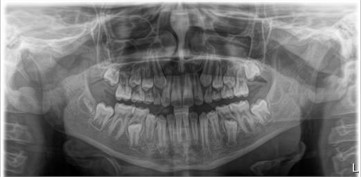
A panoramic radiography confirmed the patient’s stage of dental development and the presence of mixed dentition in second phase.
The clinical records included extraoral photographs, frontal and lateral intraoral photographs, maxillary and mandibular occlusal views, and panoramic radiography.
Fig. 1Initial extraoral photographs: a) facial asymmetry, head tilt to the right with mandibular deviation to the left; b) the working side is the left side, and this side of the face is shorter, with reduced vertical dimension at the occlusal plane; c) milder concave profile at rest; d) more pronounced concave profile upon smiling
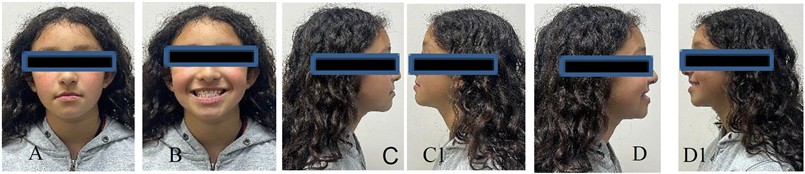
Fig. 2Initial intraoral photographs show: a) right occlusion; b) frontal occlusion; c) left occlusion; d) maxillary occlusion; e) mandibular occlusal
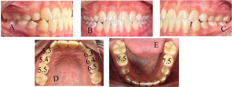
Fig. 3Initial panoramic radiography, mixed dentition sample, second phase
3. Diagnostic and therapeutic approach
The diagnostic approach considered both the static occlusal relationship and the functional characteristics of mandibular movement and chewing, with special attention to facial asymmetry, mandibular deviation, inclination of the occlusal plane, and the predominant working side. The therapeutic objective was not only to correct the previous dental relationship, but also sought to eliminate the functional interference associated with the previous cross bite, reorganize the occlusal plane, improve the previous guide and favor a more balanced masticatory pattern. Due to the eruption of the permanent upper central and lateral incisors, the conventional application of Direct Flat Tracks on these teeth was limited. Therefore, an individualized adaptation was chosen, using the remaining primary teeth as support for resin increments.
Treatment consisted of:
– Centering the upper and lower dental midlines.
– Establishing the appropriate vertical relationship to allow the anterior crossbite to be released.
– Applying light-curing resin increments to selected remaining primary teeth.
– Performing selective occlusal adjustments when necessary.
– Monitoring the relationship between the resin increments and the erupting permanent teeth.
– Providing tongue-posture reeducation when indicated.
– Guiding the patient toward an alternating bilateral masticatory pattern.
– Periodically modifying and wearing the resin increments to avoid interference with the eruption of permanent teeth.
The procedures and subsequent clinical controls were recorded through the digital platform and evaluated by the specialist responsible for the case.
4. Application of light-curing resin increments
The resin increments were applied to selected remaining primary teeth according to the functional requirements of the case.
The clinical objective was to create a controlled occlusal contact that would help release the anterior crossbite and modify the functional relationship between the maxilla and mandible.
The upper and lower dental midlines were centered before the resin was applied. A bite registration was obtained when necessary to determine the space available for the resin increments.
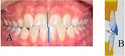
In the upper primary canine region, increments were applied to teeth 5.3 and 6.3. The resin was applied in a veneer-like configuration on the vestibular surface and extended toward the gingival direction to create an inclined functional plane. Additional resin was placed toward the vestibular-incisal region to provide sufficient height for a vis-vis relationship at the level of the primary canines. Additional resin increments were also applied to selected primary molars according to the functional requirements identified during treatment. The intervention was conservative and designed to preserve the eruption path of the permanent teeth.
Fig. 4a) Frontal view of occlusion, with deviation from the midline (Photo by the authors); b) diagram of primary maxillary canine photocuring resin increments [5.3 and 6.3]. Diagrams show the planned position and inclination of resin increments to establish the intended functional contact. Source: Diploma of Functional maxillary orthopedics Dr. Santiago Araya
See if the tongue is located between the dental arches. If so, language re-education exercises are recommended [put the tongue on the palate 5 times after each meal].
Center Upper and lower midlines (Fig. 4(a)).
Observe the space to be filled with the resin in 5.3 and 6.3 Apply intricate resin increments of type shell vestibular and gingival direction forming an inclined plane in 5.3-6.3 (Fig. 4(b)).
Also apply resin increments on the vestibular incision edge, giving enough height to achieve the previous uncrossing.
Fig. 5a) Clinical instructions and b) diagrams illustrate the planned application of photocuring resin increments in the upper and lower primary molars
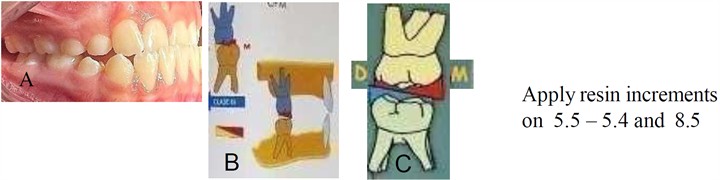
Center the upper and lower dental midlines [to observe the space to be filled with photocuring resin increments]. Make the bite registration with the available material [heavy silicone or utility wax]. Remove one of the bites from one side, observing the space to be filled with the resin increments in 5.5- 5.4- 8.5.
Fig. 6a) Clinical instructions and b) diagrams showing the planned distribution of photocuring resin increment in primary molars
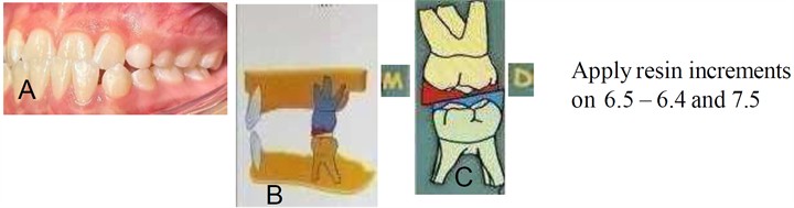
Center the upper and lower dental midlines [to observe the space to be filled with photocuring resin increments]. Record the bite with the available material [heavy silicone or utility wax]. Remove one of the bites from one side, observing the space to be filled with the resin increments at 6.5 - 6.4 - 7.5.
5. Functional reeducation
Functional rehabilitation was an integral part of the treatment. The patient was instructed to develop an alternate bilateral masticatory pattern, with alternating feeding from side to side in a balanced manner. The recommended chewing pattern included:
– alternating bilateral chewing; an initial previous bite.
– lateral mandibular movements.
– efficient shredding and grinding of food.
The patient was also encouraged to consume food with appropriate mechanical consistency, especially hard, fibrous and dry, according to the principles of neuro occlusal rehabilitation [2].
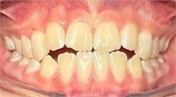
Fig. 7Initial intraoral photographs showing the previous cross bite which compromises the four permanent incisor jaws before treatment
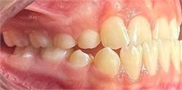
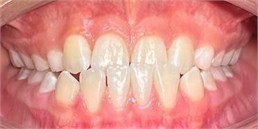
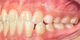
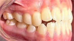
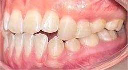
Fig. 8Clinical photographs showing the installation of photocured resin increments on the remaining primary teeth, the same day of photography
6. Eruptive control and management
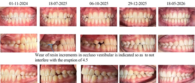
The patient was monitored periodically from November 2024 to May 2026. During follow-up, a progressive improvement of the previous cross bite was observed. The occlusal plane became more balanced and the mandibular functional pattern improved.
As the permanent teeth continued to erupt, the resin increments were progressively modified.
The occlusive-vestibular wear of the resin was carried out when necessary to avoid interference with the eruption of permanent teeth, especially premolars and canines.
The resin increments were reduced and eventually eliminated as the permanent teeth erupted, and the occlusal relationship was established.
In clinical control, an improvement was also observed in the previous guide and a progressive transition from the initial unilateral masticatory pattern to the bilateral alternation of chewing.
No functional discomfort related to resin increments was reported.
Fig. 9Clinical control of photocured resin increments from November 2024 to May 2026. Progressive occlusive-vestibular palatine wear was performed when necessary to avoid interference with the eruption of permanent teeth
7. Results
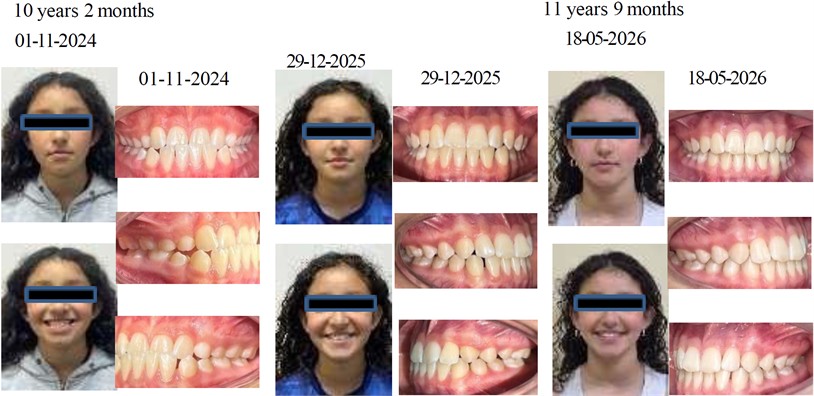
The clinical case showed a progressive correction of the previous cross bite involving the four superior permanent incisors after photocuring resin increments were applied to the remaining primary teeth.
The main clinical changes observed during follow-up were:
– Correction of the previous cross bite.
– Improvement of the occlusal plane.
– Improvement of the previous guide.
– Reduction of mandibular functional deviation.
– Progressive transition to alternate bilateral chewing.
Fig. 10Extraoral and intraoral photographs comparing the initial and final clinical conditions following treatment with light-curing resin increments
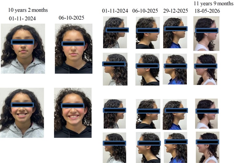
Fig. 11Photographs comparing the initial and final clinical conditions
8. Discussion
The present clinical case describes an individualized adaptation of the principles of Planas Direct Tracks within the framework of neuro-occlusal rehabilitation for the management of a functional anterior cross bite during the second phase of mixed dentition.
The main clinical difficulty was related to the four permanent upper incisors erupted. As these teeth were already present, the conventional application of Planas Direct Tracks directly to the anterior teeth was not possible. The use of the remaining primary teeth as support for photocuring resin increments provided an alternative means to modify the anomalous to functional occlusal plane while preserving the eruptive trajectory of the permanent teeth.
Treatment was based on the concept that occlusal interference can influence mandibular trajectories and masticatory function. Therefore, the therapeutic goal was not simply to move the incisors but to modify the functional conditions that contributed to the previous cross bite.
Previous reports have described the use of Planas Direct Tracks for the early correction of functional crossbites. Pereira et al. [15] reported the correction of a functional anterior crossbite using Planas Direct Tracks and emphasized the importance of differential diagnosis and elimination of occlusal interferences.
Similarly, Ciarcia et al. [16] reported successful early treatment of anterior and posterior crossbite using direct dental tracks, with clinical stability during a three-year follow-up. Although the technique used in that case followed the conventional approach, the findings support the importance of early intervention and the potential clinical relevance of adapting the principles of neuro-occlusal rehabilitation to different stages of dental development.
Lentini-Oliveira [10], however, highlighted that the scientific evidence regarding Planas Direct Tracks remains limited, with a predominance of case reports and case series and a scarcity of comparative studies. Therefore, the findings of the present case should be interpreted as clinical evidence of the feasibility of an individualized therapeutic approach rather than as evidence of generalized treatment efficacy.
The functional dimension of the intervention is also relevant. The observed improvement in the masticatory pattern occurred together with the correction of the anterior crossbite and the improvement of the occlusal plane. From the perspective of neuro-occlusal rehabilitation, the functional stimulus generated by the new occlusal condition may contribute to the reorganization of mandibular movement and masticatory function [2, 3].
The use of light-curing resin increments also provided a conservative alternative in a patient in whom the conventional application of Planas Direct Tracks was limited by the eruption stage. Periodic adjustment of the resin was essential to avoid interference with the eruption of permanent teeth.
Another relevant aspect of this case was the integration of asynchronous teleorthodontic into primary health care. The digital platform allowed clinical records to be assessed by specialists and therapeutic recommendations to be communicated to the primary care team. This approach may facilitate timely management of interceptive cases and reduce unnecessary referrals while maintaining specialist supervision [7, 11].
However, the limitations of a single clinical case must be recognized. The favorable clinical evolution observed in this patient cannot be generalized to all patients with anterior crossbite. In addition, the present report does not allow comparison with other treatment modalities or determination of long-term stability beyond the follow-up period described.
Prospective studies involving larger samples and standardized functional and occlusal assessments are necessary to determine the reproducibility, stability, and comparative effectiveness of this therapeutic adaptation.
9. Conclusions
For this clinical case, the use of photocured resin increments in the remaining primary teeth, combined with selective occlusal adjustments and functional rehabilitation, was associated with favorable clinical changes in a previous cross bite involving the four superior permanent incisors.
The intervention was carried out within the principles of neuro occlusal rehabilitation and allowed a progressive improvement of the anterior occlusal relation, the occlusal plane and the masticatory pattern, while preserving the eruption of the permanent teeth. The case also demonstrates the feasibility of integrating asynchronous teleorthodontic support specialized in primary health care for the management of interceptive occlusal problems.
As this is a unique clinical case, these findings should be interpreted with caution. More clinical studies are needed to determine whether this approach provides reproducible and stable results in a larger population.
References
-
W. A. Simões, P. Valerio, and D. Duarte, Functional Orthopedics of the Maxilla: Research and Clinical Excellence. São Paulo, Brazil: Napoleão, 2020.
-
W. A. Simões, Functional Orthopedics of the Jaws Through Neuroocclusal Rehabilitation. Madrid, Spain: Editorial Médica Panamericana, 2004.
-
P. Planas, Neuro-occlusal rehabilitation. Medellín, Colombia: Amolca, 2008.
-
S. Silveira, P. Valerio, and A. J. Machado Júnior, “The law of minimum vertical dimension: evidence for improvement of dental occlusion,” European Journal of Dentistry, Vol. 16, No. 2, pp. 241–250, Oct. 2021, https://doi.org/10.1055/s-0041-1732950
-
O. Quirós Álvarez, C. J. Quirós, and O. J. Quirós, Use of Direct Tracks for the Early Correction of Skeletal Malocclusions. Use of the Bite Track Maker. Bogotá, Colombia: Amolca, 2022.
-
R. R. D. Santos, A. J. Isper Garbin, and C. A. Saliba Garbin, “Early correction of malocclusion using Planas direct tracks,” Case Reports in Dentistry, Vol. 2013, pp. 1–4, 2013, https://doi.org/10.1155/2013/395784
-
“Orthodontics cell,” Ministerio de Salud de Chile, Portalsaluddigital.minsal.cl, 2026.
-
A. J. I. Garbin, B. Wakayama, T. A. Saliba, and C. A. S. Garbin, “Intervention in posterior crossbite using Planas direct tracks: seven-year follow-up case report,” Revista Cienciasde La Salud, Vol. 18, No. 2, pp. 1–10, 2020.
-
C. I. S. A. Furtado, R. L. Silva, K. B. Souza, and N. M. Fontes, “The use of Planas direct tracks in the treatment of functional crossbites,” in Jornada Odontológica Dos Acadêmicos Da Católica, Vol. 10, 2022.
-
D. A. Lentini-Oliveira, “Planas direct tracks to treat functional crossbites in children: scientific evidence,” Jaw Functional Orthopedics and Craniofacial Growth, Vol. 2, No. 2, pp. 24–31, Nov. 2022, https://doi.org/10.21595/jfocg.2022.22739
-
“Telehealth and remote care: guidelines for the care network,” Ministerio de Salud de Chile, Interconsulta.minsal.cl, 2026.
-
C. M. Borțun and L. C. Rusu, “Recording of mastication angles by Planas’s laws,” Stomatology Education Journal, Vol. 1, No. 2, pp. 86–91, 2014.
-
L. A. Almeida-Junior, M. M. Polimeno, A. P. Tortelli, and V. Medau, “Neuroclusal rehabilitation with Planas direct tracks in the treatment of anterior crossbite – literature review,” Jaw Functional Orthopedics and Craniofacial Growth, Vol. 4, No. 1, pp. 39–43, 2024, https://doi.org/10.21595/jfocg.2024.23837
-
G. R. Matos, R. R. Neto, A. J. M. Júnior, and R. B. B. Junior, “Influence of the inclination of the incisal edge of Planas direct tracks on deciduous dentition with anterior crossbite: finite-elements study,” European Journal of Dentistry, Vol. 16, No. 3, pp. 528–535, 2021, https://doi.org/10.1055/s-0041-1735435
-
A. P. S. Pereira, G. B. R. Prestes, N. Sarmento, A. L. T. Dutra, V. S. L. Braga, and C. I. O. Carvalhal, “Pista direta planas para tratamento de mordida cruzada anterior funcional – relato de caso,” Orthodontic Science and Practice, Vol. 14, No. 53, pp. 56–61, 2021, https://doi.org/10.24077/2021;1453-5661
-
A. C. C. D. M. Ciarcia, M. F. X. B. Cavalcanti, F. Diomede, M. P. S. M. Pugliese, S. R. G. Olivan, and O. M. Altavista, “Early treatment of a preschool child’s crossbite using direct dental tracks, three-year follow-up – clinical case report,” Brazilian Journal of Implantology and Health Sciences, Vol. 8, No. 2, pp. 1195–1210, 2026, https://doi.org/10.36557/2674-8169.2026v8n2p1195-1210
About this article

The authors have not disclosed any funding.
The datasets generated during and/or analyzed during the current study are available from the corresponding author on reasonable request.
Ruth Del Campo Yáñez: conceptualization, methodology, investigation. Carolina Romano Cornejo: clinical work, composing original draft. Paula Pino Vásquez: clinical work. Santiago Araya Zavala: clinical supervision, review and editing, supervision of clinical treatment.
The authors declare that they have no conflict of interest.
Informed consent of the patient and her legal guardian was obtained for the treatment and publication of this clinical case, including the use of clinical photographs and radiographic records. The study was conducted in accordance with the ethical principles of the Helsinki

